Carl Zeiss Meditec AG (Germany)
Diagnostic technology
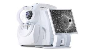
ZEISS OCT
Gold-standard ZEISS OCT for non-invasive 3D retinal and anterior segment imaging — the definitive tool for detecting, diagnosing, and monitoring sight-threatening eye diseases.
Overview
How it works
The ZEISS Cirrus HD-OCT is a gold-standard, non-invasive imaging system used at Peshawar Eye Hospital to produce high-resolution 3D images of the retina and anterior segment. It enables early detection and monitoring of conditions such as glaucoma, diabetic retinopathy, and age-related macular degeneration, while also assessing corneal thickness and the anterior chamber for comprehensive eye evaluation.
Key benefits
- Non-invasive, instant high-resolution 3D cross-sectional imaging of all retinal layers
- Detects silent eye diseases before any visual symptoms appear
- Microscopic retinal layer analysis with micron-level precision
- Tracks anti-VEGF treatment response with objective quantitative measurements
- Anterior segment OCT module for corneal thickness and angle imaging
- Trusted globally as the clinical standard for retinal and glaucoma management
What we use it for
- Macular Disease Detection (AMD) — Provides cross-sectional 3D images of all retinal layers, detecting sub-retinal fluid, drusen, and pigment epithelial detachments in age-related macular degeneration before central vision is affected.
- Glaucoma Screening & Monitoring — Measures optic nerve fibre layer (RNFL) thickness with reference to normative databases, enabling early detection of glaucomatous nerve damage and objective monitoring of disease progression.
- Diabetic Retinopathy Assessment — Quantifies diabetic macular oedema (DMO) with retinal thickness maps, guides anti-VEGF injection timing, and confirms treatment response at follow-up appointments.