
Diagnostic technology
Visual Field Perimetry
Computerised perimetry mapping the entire peripheral and central visual field — the gold standard for early glaucoma detection and neurological visual pathway assessment.
Overview
How it works
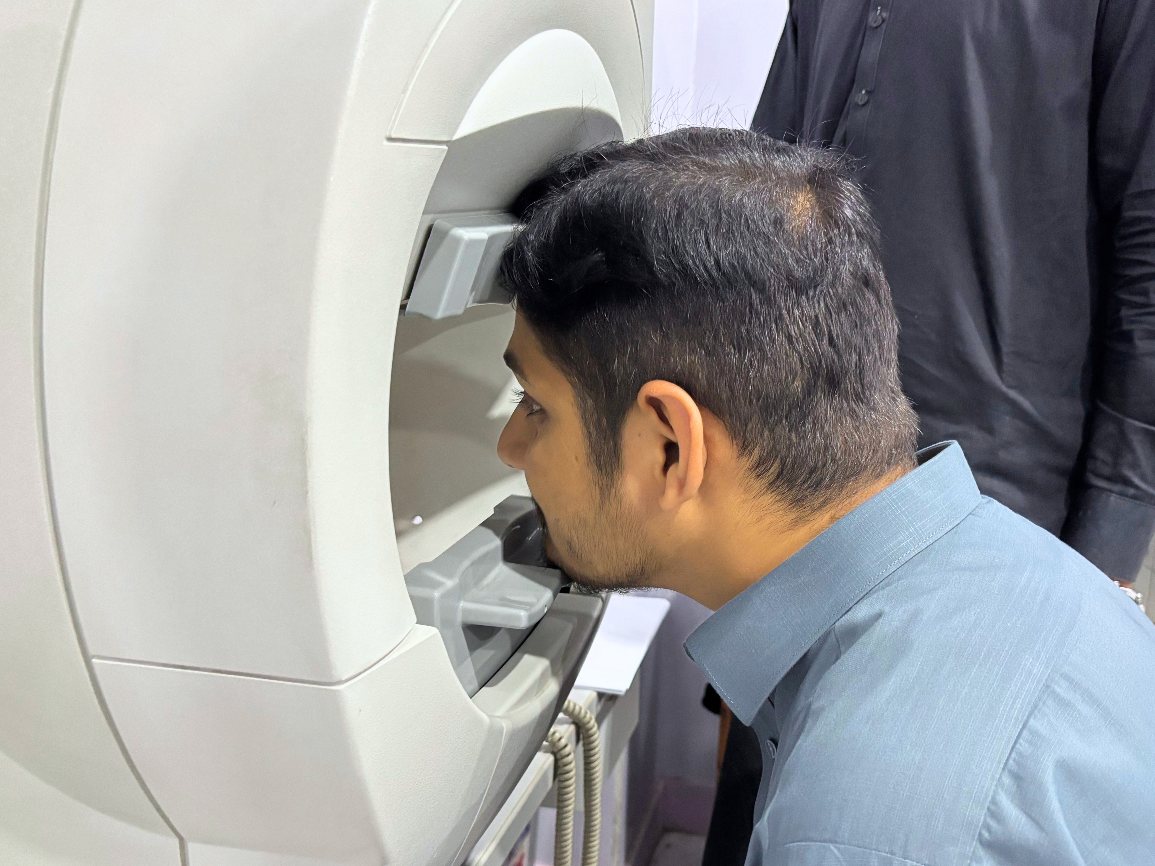
Visual Field Perimetry at Peshawar Eye Hospital provides computerised mapping of the entire central and peripheral visual field — the gold-standard investigation for early glaucoma detection, neurological visual pathway assessment, and occupational vision fitness. The system, manufactured by Haag-Streit and Carl Zeiss Meditec, uses the gold-standard SITA (Swedish Interactive Threshold Algorithm) to test hundreds of points across the visual field efficiently and accurately, producing a detailed sensitivity map that identifies scotomas — areas of reduced or absent vision — caused by optic nerve damage. These defects are detectable years before the patient notices any visual change, making perimetry a critical tool for catching glaucoma at a stage when treatment can still preserve sight. Beyond glaucoma, characteristic field defect patterns such as hemianopia or arcuate scotomas allow neurologists and ophthalmologists to localise lesions along the entire visual pathway. The GPA (Guided Progression Analysis) module automatically compares serial tests over time to detect statistically meaningful change.
Key benefits
- Comprehensive mapping of the entire peripheral and central visual field in one test
- Detects glaucomatous visual damage years before the patient notices any vision loss
- Completely non-invasive and painless — No drops, No contact, No radiation
- Progression analysis compares serial tests to detect statistically significant change
- Gold-standard SITA (Swedish Interactive Threshold Algorithm) for accurate fast testing
- GPA (Guided Progression Analysis) provides automated change detection over time
What we use it for
- Glaucoma Screening & Progression Monitoring — Computerised static perimetry maps the sensitivity of the visual field at hundreds of test points, detecting scotomas (blind spots) caused by glaucomatous optic nerve damage long before the patient notices any visual change.
- Neurological Visual Pathway Evaluation — Characteristic visual field defect patterns — hemianopia, quadrantanopia, arcuate scotomas — help identify and localise neurological lesions affecting the optic nerve, chiasm, optic tract, or cortical visual pathways.
- Driving & Occupational Fitness Assessment — Provides objective, reproducible documentation of peripheral vision adequacy for driving licence medical requirements and occupational vision standards for pilots, police, and other regulated professions.