Diagnostic technology
Eye Ultrasound — A-Scan & B-Scan
High-frequency ocular ultrasonography that sees deep inside the eye through dense cataracts, haemorrhage, or trauma — essential for retinal diagnosis and IOL power planning.
Overview
How it works
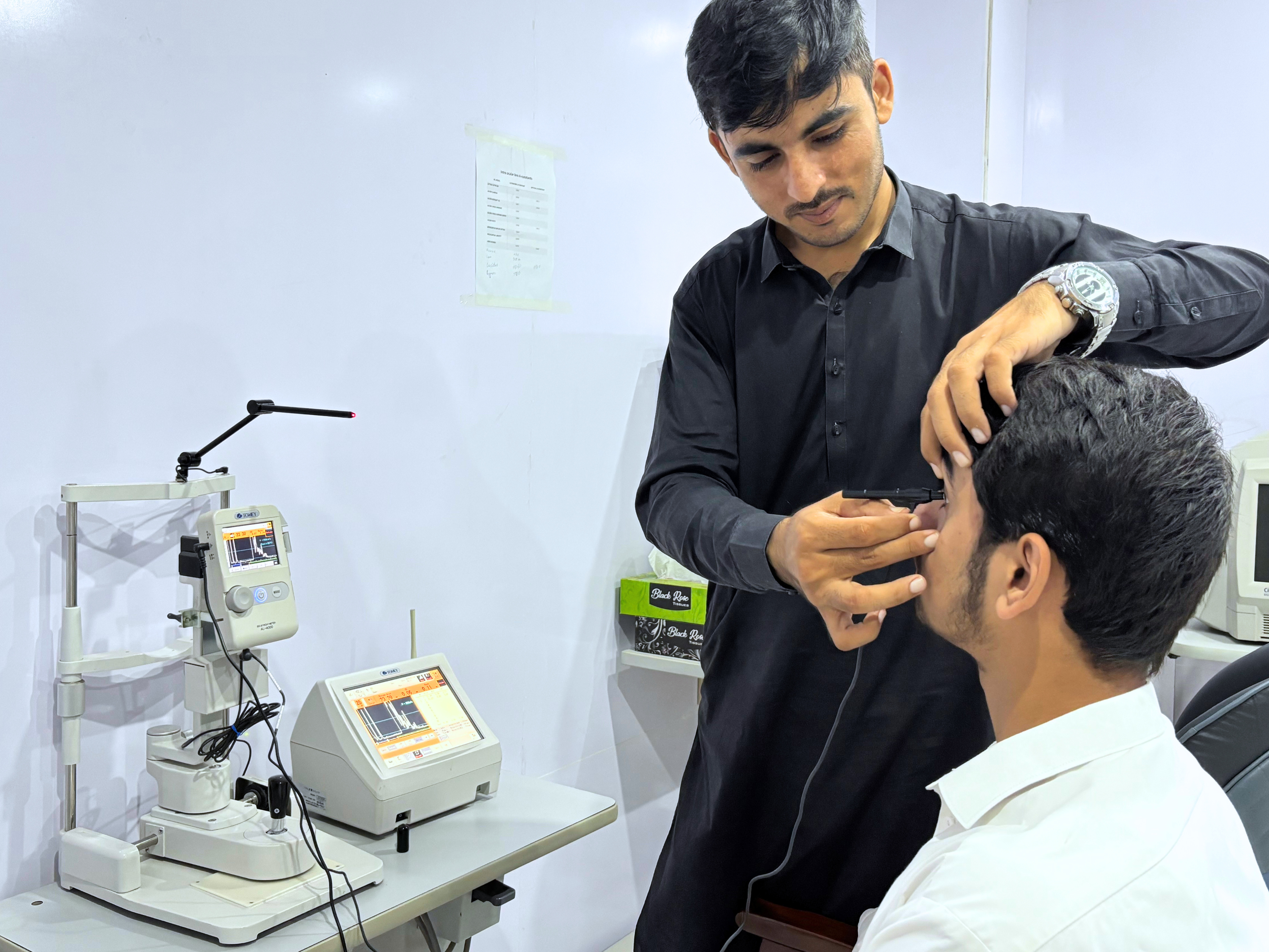
Ocular ultrasonography at Peshawar Eye Hospital uses high-frequency sound waves to image the internal structures of the eye when direct visualisation is impossible — in cases of dense cataract, vitreous haemorrhage, corneal scar, or ocular trauma. The B-Scan provides a real-time two-dimensional cross-sectional view of the entire eye, enabling rapid diagnosis of retinal detachment, posterior vitreous detachment, intraocular tumours, and the source of vitreous bleeding even when the media is completely opaque. This information is essential before proceeding with cataract surgery on an eye with no fundal view, or planning a vitreo-retinal operation. The A-Scan mode measures the precise axial length of the eye — the single most critical variable for calculating the correct IOL power in cataract surgery — ensuring patients achieve their target refraction after lens implantation. Systems are manufactured by Quantel Medical in France and Accutome in the USA, providing both diagnostic and biometric capabilities on a single device.
Key benefits
- Penetrates dense media — cataracts, vitreous blood, corneal scars — to image the posterior segment
- Rapid retinal detachment diagnosis even when direct fundoscopy is impossible
- A-Scan axial length measurement for accurate IOL power calculation
- Real-time B-Scan cross-sectional imaging of the entire eye in seconds
- Identifies intraocular tumours, choroidal lesions, and abnormal ocular swelling
- Indispensable pre-operative planning tool for complex cataract and vitreo-retinal cases
What we use it for
- Pre-Operative Dense Cataract Assessment — When a dense cataract completely blocks the ophthalmoscopic view of the retina, B-Scan ultrasound is the only way to confirm retinal attachment and identify pathology before proceeding with cataract surgery.
- Vitreous Haemorrhage Diagnosis — B-Scan imaging detects retinal detachment, posterior vitreous detachment, and the source of intraocular bleeding in real time, even when blood fills the vitreous cavity and obscures all direct visualisation.
- IOL Power Calculation (A-Scan Biometry) — Immersion A-Scan precisely measures the axial length (front-to-back distance) of the eye — the most critical variable in calculating the correct IOL power for cataract surgery to achieve the target refraction.